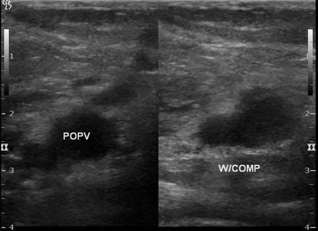
Image of a doppler ultrasound scan showing a thrombosis in the popliteal vein Source
LAST WEEK, I gave a presentation on the ultrasound diagnosis of chronic deep vein thrombosis—and it was a fascinating (though sobering) topic. While researching, I realized just how serious DVT is—something no patient wants to hear their doctor call "interesting." Because in medicine, an "interesting case" often means the patient is in real trouble.

DVT isn’t something to take lightly. It can happen to anyone, anywhere, anytime—though certain risk factors increase the odds. In the U.S. alone, around 500,000 tests are ordered annually to rule out DVT, and over 100,000 deaths may be linked to it (directly or indirectly). Many of these deaths go unautopsied, but pulmonary embolism—a top cardiovascular killer after stroke and coronary artery disease—often stems from untreated DVTs.
So, What Exactly Is DVT?
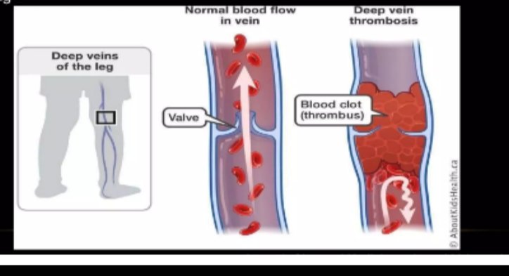
In simple terms, DVT is abnormal blood clotting in the deep veins, usually in the legs (like the femoral veins). These veins rely on muscle movement to push blood back to the heart. If you’re inactive for too long, blood stagnates, raising the risk of clots.
That’s why sitting for extended periods is so dangerous. Movement is key! Fun fact: Your body has a "second heart"—the gastrocnemius muscle (the calf muscle behind your knee). When you walk, it contracts, squeezing veins and arteries to pump blood upward, just like the heart. No movement? Blood pools, and clots form.
The 3 Root Causes of DVT (Virchow’s Triad)
- Endothelial damage – Injury to blood vessel walls triggers clotting, even without major bleeding.
- Stasis – Poor blood flow (e.g., from immobility) lets blood stagnate and clot.
- Hypercoagulability – Conditions like cancer, pregnancy, birth control pills, or post-surgery states make blood clot too easily.

What Does DVT Look Like on Ultrasound?
Clots appear as obstructive plaques in veins, varying in type:
- Floating (free-moving)
- Occlusive (fully blocking the vein)
- Donut-shaped (partial recanalization)
- Marginal (clinging to vein walls)
Radiologists must identify the type, location, and size to guide treatment—whether clot removal or medication. Gentle scanning is crucial; aggressive pressure can dislodge clots, causing a fatal pulmonary embolism.
The Bottom Line
DVT can lead to leg amputation (from blocked blood flow) or sudden death (if a clot reaches the lungs). Stay active, walk regularly, and avoid long periods of sitting. Your "second heart" needs exercise too!
Take care—and keep moving. 💙
Dont stop Moving.
Images taken from my slide which are sourced from Source Link